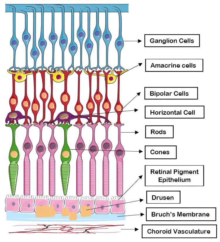
Figure 2. Retinal cell layers: drusen between RPE and BrM.
Parts of the figure were drawn using pictures from Servier Medical Art. Servier Medical Art by Servier is licensed under a Creative Commons Attribution 3.0 Unported License (https://creativecommons.org/licenses/by/3.0/).
RPE, retinal pigment epithelium; BrM, Bruch's membrane

