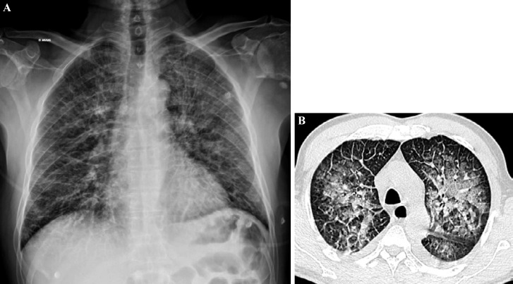
Figure 15.
Patient âgé de 55 ans admis en réanimation pour prise en charge d'un syndrome pulmonaire à hantavirus compliqué d'une défaillance multiviscérale (respiratoire, rénale et hématologique) (crédit photo: M. Zappa)
Légende: La radiographie pulmonaire (A) et le scanner thoracique (B) (fenêtre parenchymateuse passant par les lobes supérieurs) montrent des condensations alvéolaires et verre dépoli en plage prédominant dans les régions centrales associés à des lignes septales. Évolution favorable sous traitement symptomatique de réanimation et antibiothérapie.
55-year-old patient admitted in intensive care unit for management of a pulmonary hantaviral syndrome complicated by multivisceral failure (respiratory, renal and hematological) (photo credit: M. Zappa)
Chest X-ray (A) and CT scan (B) (parenchymal window through the upper lobes) show alveolar condensations and ground glass predominantly in the central regions associated with septal lines. Favorable evolution under symptomatic resuscitation treatment and antibiotic therapy.

