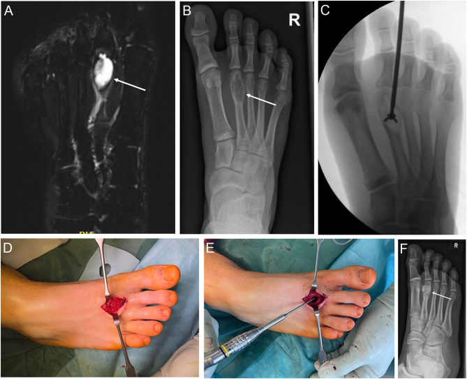
Figure 5.
(A) MRI of an aneurysmal bone cyst with high tumour signal intensity seen on the T2-weighted imaging and eccentric lucent bone lesion on x-ray (B) in a 17-year-old male patient with aneurysmal bone cyst confirmed by biopsy (C). Surgical treatment consisted of enucleation, curettage, milling the cyst cavity (D, E) and defect filling with allogeneic cancellous bone graft (F).

 This work is licensed under a