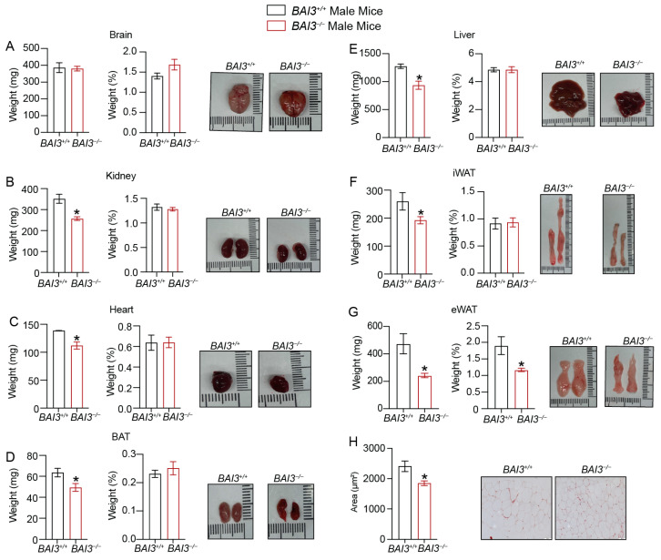
Figure 3.
Assessment of weight of tissues obtained from BAI3+/+ and BAI3−/− male mice. (A–G) Tissues were harvested from eight-week-old BAI3+/+ and BAI3−/− male mice. The absolute tissue weight (left), normalized tissue weight to the total BW (middle), and gross morphology (right) for the brain, liver, kidney, heart, eWAT, iWAT, and BAT are presented (n = 8–10). (H) H and E staining of eWAT for determining adipocyte size. Data are expressed as mean ± SEMs, * p < 0.05.

