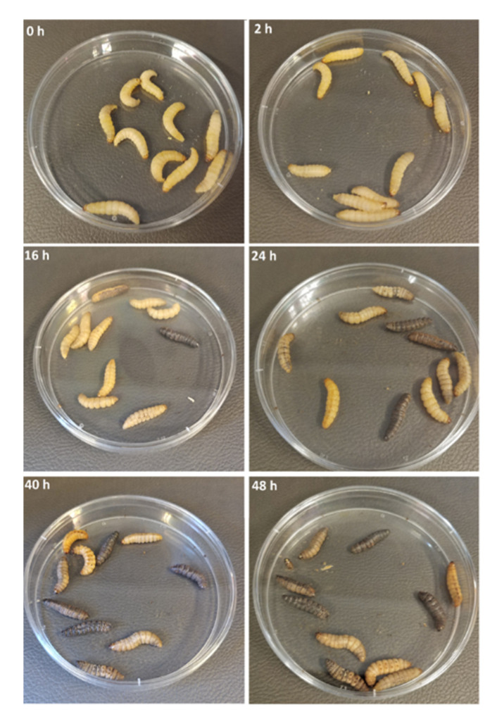
Figure 3.
Images of larvae infected with gonococci. The images show the development of melanisation, i.e., the observed blackening of the larval body as a marker of larval death with time. © Dijokaite, Humbert, Borkowski, La Ragione, Christodoulides. Reproduced from Dijokaite, A.; Humbert, M.V.; Borkowski, E.; La Ragione, R.M.; Christodoulides, M. Establishing an invertebrate Galleria mellonella greater wax moth larval model of Neisseria gonorrhoeae infection. Virulence 2021, 12, 1900–1920, https://doi.org/10.1080/21505594.2021.1950269 [291].

