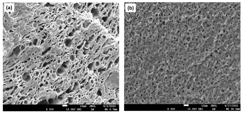
Figure 1.
Porous structures of fractured biopolymer-based scaffolds. Scanning electron microscopy (SEM) imaging technique was applied for the analysis of the scaffolds’ cross-sections. (a) An image depicting the porous architecture of the matrix. Both larger, as well as smaller pores, are displayed, which are distributed in an inhomogeneous manner; (b) observation of the multiple micropores with a homogenous distribution, creating the inner environment for cell attachment, proliferation, and migration, as well as differentiation.

