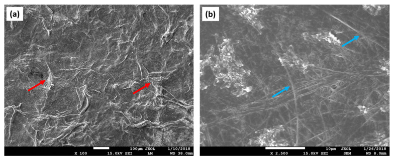
Figure 3.
SEM images revealing the surface topography of the collagen scaffold. (a) Collagen fibers (red arrows) create the rough surface of the scaffold. Depicted design enlarges the overall surface of the matrix, thus providing a larger area for the cell attachment; (b) a detailed analysis of the scaffold’s architecture displays collagen fibers oriented in an unaligned manner (blue arrows).

