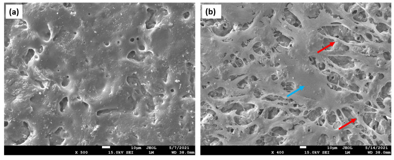
Figure 5.
PLA-based scaffold. (a) Surface analysis of the PLA scaffold engineered by the pressing technique (reference sample). SEM image displaying smooth, homogenous area for the cell attachment; (b) PLA-based scaffold seeded with human fibroblasts (blue arrow). After 7 days of incubation, a dense cellular layer on the scaffold surface could be observed. Multiple filopodia (red arrows) are also detected, proving efficient attachment to the scaffold surface. The presented findings demonstrate good scaffold-cell interactions and determine good biocompatibility of the material.

