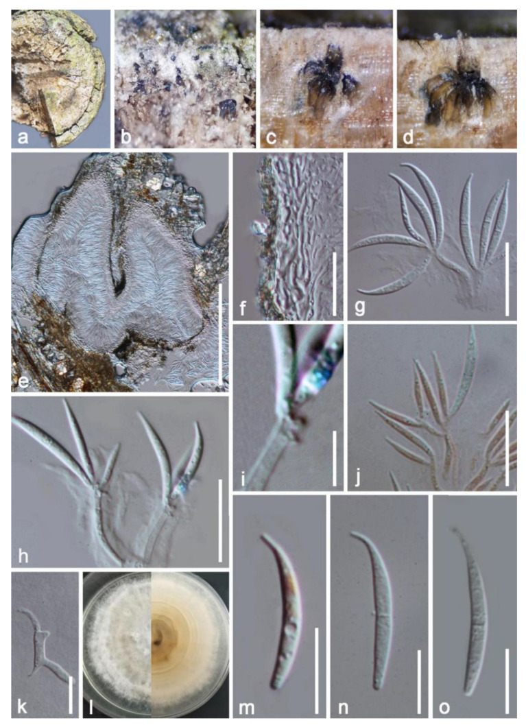
Figure 10.
Delonicicola siamense (HKAS 122662). (a,b) The appearance of the natural substrate; (c–e) cross section of conidiomata; (f) conidiomata wall; (g,h) conidia with conidiophore; (j) conidiophore and conidia stained by Congo red reagent; (i) close-up of conidiogenous cells; (k) germinated conidium; (l) colonies on PDA; (m–o) Conidia. Scale bars: (e) = 200 μm; (f,g) = 20 μm; (j,h) = 15 μm; (m–o) = 10 μm; (i) = 5 μm.

