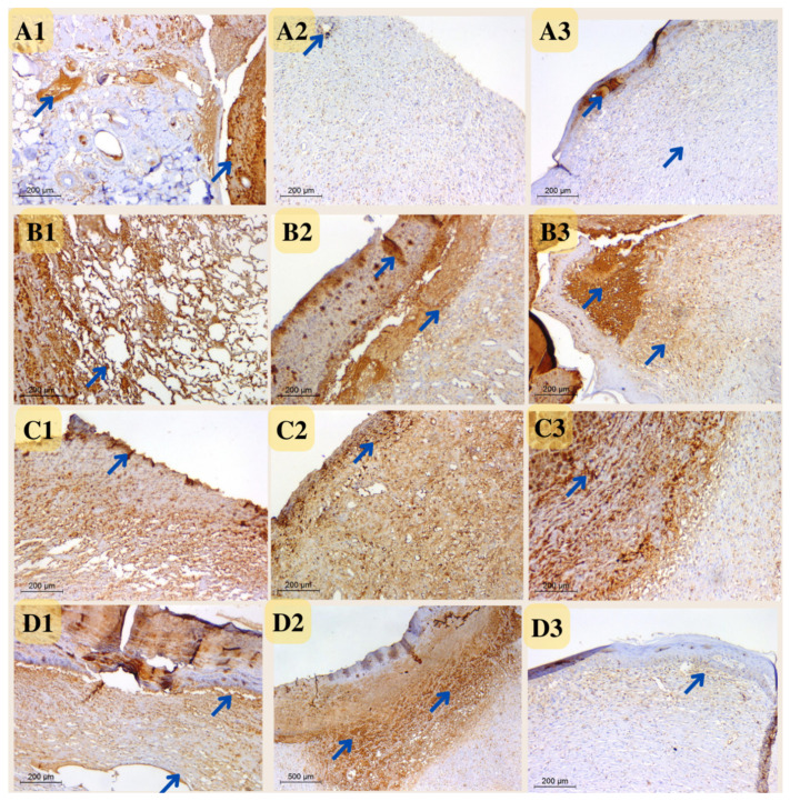
Figure 15.
Photomicrograph of wound tissue sections stained with CD31 expression as angiogenesis factor in rat wounds on days 4 (coded number 1), 8 (coded number 2) and 12 (coded number 3) (×200). (A1–A3) control group; (B1–B3) positive control group; (C1–C3) Eucalyptus oil/cellulose acetate nanofiber-treated group; (D1–D3) nano-chitosan/Eucalyptus oil/cellulose acetate nanofiber-treated group. Arrows indicate the stained vesicles.

