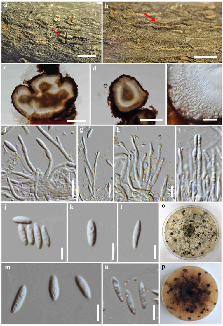
Figure 7.
Diaporthe samaneae (CMUB39997, holotype). (a,b) Conidiomata on host substrate (indicated with the red arrow). (c,d) Section through conidiomata. (e) Conidiomatal walls. (f–i) Conidiogenous cells giving rise to conidia. (j–n) Alpha conidia. (o,p) Colonies on PDA, (o) from above and (p) from reverse. Scale bars: (a,b) = 500 μm, (c,d) = 200 μm, (e) = 20 μm, (f–i) = 10 μm, and (j–n) = 5 μm.

