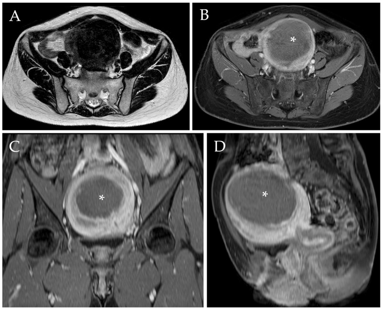
Figure 4.
Post-procedural MRI of the same patient. Axial T2-w image (A) demonstrated a reduction of the diameter of the voluminous myoma after UAE, which now measures approximately 90 mm (i.e., a diameter reduction of 35.7%). Axial, sagittal, and coronal post-contrast T1-w images with fat saturation (B–D) confirmed the diameter reduction and revealed the presence of a wide intra-tumoral necrotic area (white asterix).

