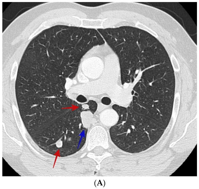
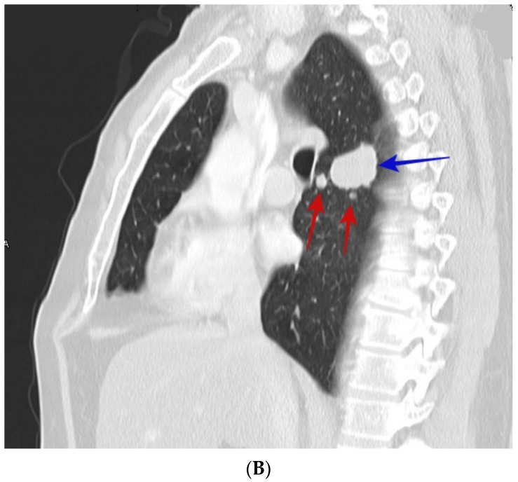
Figure 1.
(A) CT scan shows smooth right upper lobe lung nodule (blue arrow) and satellite nodules (red arrows). The patient was a 50-year-old male who presented with incidental lung nodules. CT guided biopsy showed coccidioidomycosis. (B) shows sagittal section of the right upper lobe nodule (blue arrow) and satellite nodules (red arrows) of the same patient in image Figure 1A.


