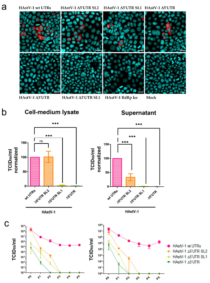
Figure 4.
Compromised viral particle assembly and release of HAstV-1 UTR deletion mutants. (a) Immunofluorescence images of CaCo-2 cells infected with supernatants of transfected BSR-T7 samples. The HAstV-1 capsid protein is stained red, and nuclei are stained blue. Scale bar = 20 µm. (b) Titration of HAstV-1 5′ UTR mutants (after BSR-T7 cell transfection = P0) on CaCo-2 cells. The titers (quantified as the 50% tissue culture infectious dose (TCID50)/mL) were normalized to those of wt HAstV-1. Data for the 3′ UTR and HAstV-1 RdRp ko mutants are not shown because these mutants did not infect CaCo-2 cells. The error bars indicate standard deviations from three independent experiments. *** p < 0.001. (c) Virus titers of HAstV-1 mutants on CaCo-2 cells in sequential passages (n = 5). P0, after BSR-T7 cell transfection; P1–P5, after CaCo-2 cell infection (n = 2), ns = nonsignificant.

