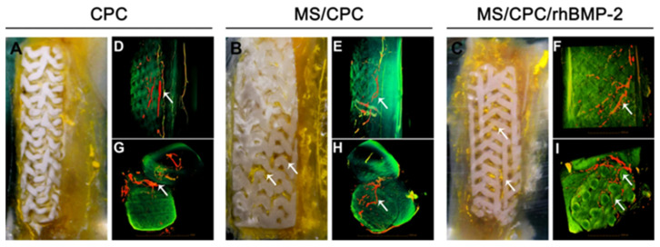
Figure 2.
(A–C): Digital camera photographs of PMMA-embedded blocks from longitudinal sections and 3D reconstructed μCT images of blood vessels from (D–F) side view and (G–I) top view of CPC, MS/CPC, and MS/CPC/rhBMP-2 scaffolds after 4 weeks of implantation. White arrow: newly formed blood vessels [77].

