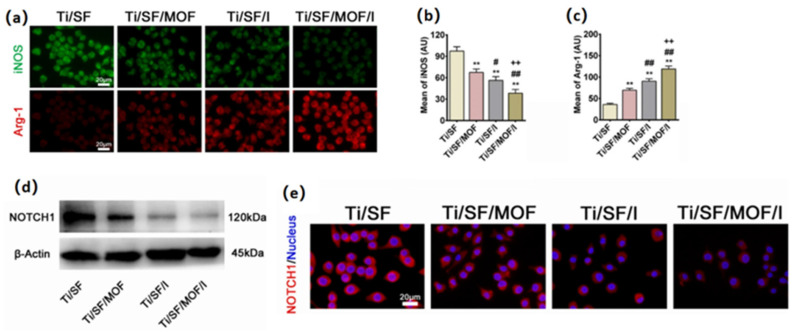
Figure 10.
(a) Immunofluorescent staining of Raw264.7 cells after cultured for 4 days. (b,c) Quantitative analysis of iNOS and Arg-1. (d) The expression of Notch1 was detected by Western blotting. (e) Immunofluorescent staining of Notch1. (n = 3; # represent p < 0.05 when compared with Ti/SF, Ti/SF/MOF and Ti/SF/I, respectively; **, ## and ++ represent p < 0.01) [80].

