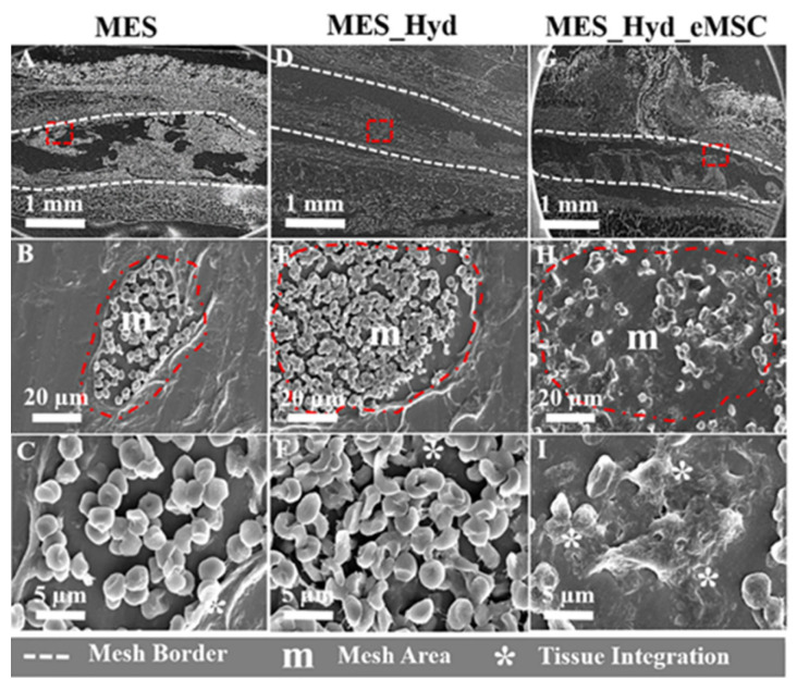
Figure 12.
Fate of meshes after 1 week of in vivo implantation. SEM images show cross-sections of (A–C) MES (D–F) MES_Hydrogel and (G–I) MES_Hydrogel_eMSC constructs (within the red dashed area) 1 week after implantation in NSG mice; lower panels (C,F,I) show the morphology of reticular fibers (m), their interaction with the host tissue integration (white*) and the formation of new ECM [122].

