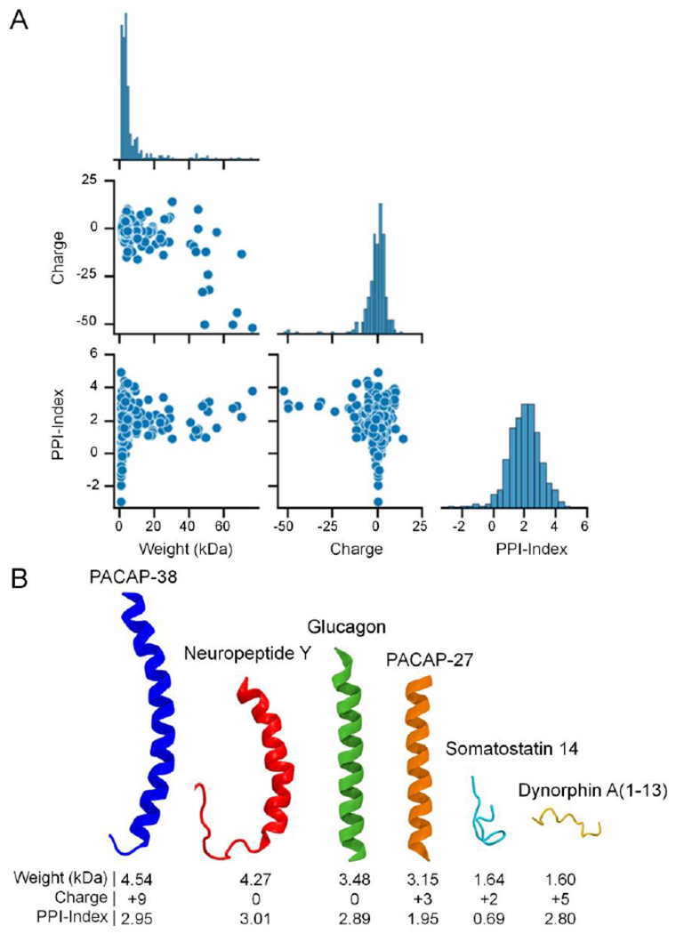
Figure 3.

The diverse physicochemical properties of neuropeptides. (A) Pair plot comparing the molecular weight (kDa), theoretical net charge at a physiologic pH of 7.4, and the Potential Protein Interaction Index (PPI-Index), a predictor of a polypeptide’s propensity to bind other proteins/receptors,49 for all 283 human neuropeptides in the NeuroPep database.48 The properties were estimated from the peptide sequences using the peptides.py package (https://github.com/althonos/peptides.py).50 Note that the diagonal edge of the pair plot shows the distributions of each property. (B) A selection of human neuropeptide structures collected from the RCSB Protein Data Bank51 and AlphaFold Protein Structure Database52, 53 highlighting the diversity of neuropeptide structure and physicochemical properties, including the 38 amino acid variant (blue) of pituitary adenylate cyclase-activating peptide (PACAP), Neuropeptide Y (red), glucagon (green), the 27 amino acid variant of PACAP (orange), Somatostatin 14 (light blue), and Dynorphin A (1-13) (yellow). The neuropeptide structures were rendered using the Visual Molecular Dynamics (VMD) software.54
