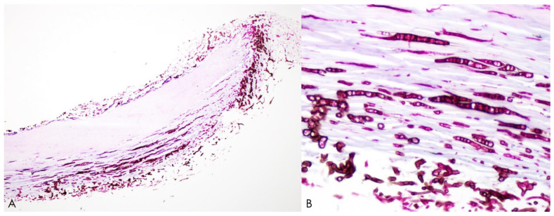
Figure 3.
Histopathological analysis of nail clippings from Neoscytalidium dimidiatum-induced onychomycosis using periodic acid–Schiff staining. Microscopic examination at low power with the 4× objective (A) and high power with the 40× objective (B) revealing black-brown fungal hyphae invading the nail plate.

