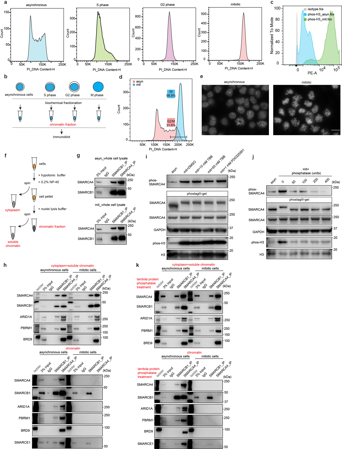
Extended Data Figure 1. Cell cycle analysis and subcellular SWI/SNF interactions in asynchronous and mitotic mouse ES cells.

a, Representative flow cytometry analysis showing DNA contents of asynchronous mouse ES cells and cells synchronized at S, G2, and mitotic phase. b, Schematic for extraction of chromatin fraction from asynchronous mouse ES cells and populations synchronized at S, G2, and mitosis. c, Representative flow cytometry showing the distribution of phosphorylated Serine 10 of histone H3 between asynchronous and synchronized mitotic mouse ES cells. d, Representative flow cytometry analysis showing DNA contents and purity of synchronized mitotic mouse ES cells. e, Representative DAPI staining confirming the high purity of synchronized mitotic cells. Scale bar: 10 μm. f, Schematic showing the isolation of cytoplasm, soluble chromatin, and chromatin fractions from mouse ES cells. g, Immunoprecipitation (IP) of SMARCA4 and SMARCB1 in whole cell lysate from asynchronous (top) and mitotic (bottom) mouse ES cells. h, IP of SMARCB1 and SMARCA4 in the cytoplasmic+ soluble chromatin fractions (top), and chromatin fraction (bottom) isolated from asynchronous and mitotic mouse ES cells. i, Immunoblots in whole cell lysate of asynchronous (lane 1) and mitotic (other lanes) mouse ES cells treated with TBB (CK2 inhibitor) and PD0325901 (ERK1 inhibitor) for 6 hours, as indicated. Samples were separated either in NuPAGE™ 4 to 12% Bis-Tris gels or in the case of the labeled SMARCA4 sample in Phos-tag™ (50 μmol/L) precast gels. j, Immunoblot in whole cell lysate of asynchronous (lane 1) or mitotic (other lanes) mouse ES cells. Lysates were treated with increasing doses of lambda protein phosphatase as indicated. Samples were separated in NuPAGE™ 4 to 12% Bis-Tris gels or in the case of the labeled SMARCA4 sample in Phos-tag™ (50 μmol/L) precast gels. k, IP of SMARCB1 and SMARCA4 in the lambda protein phosphatase treated cytoplasmic+ soluble chromatin fractions (top), and chromatin fraction (bottom) isolated from asynchronous and mitotic mouse ES cells. Data are representative of two (a, c, d, e, h, i, j, k) and three (g) independent experiments.
