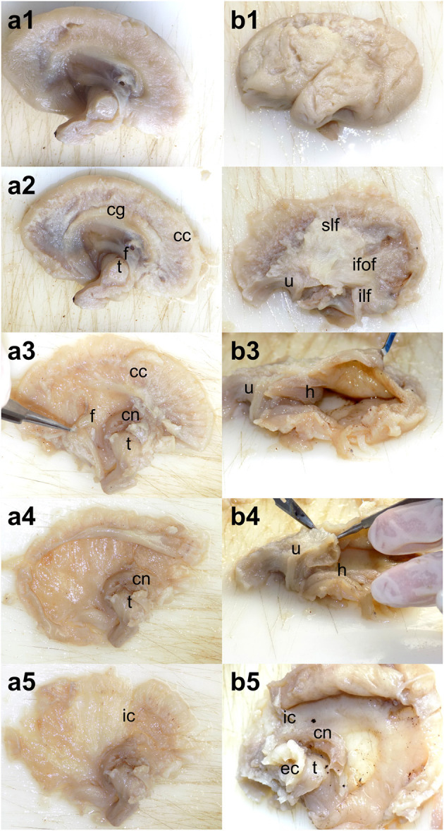
Figure 1.

Dissection steps adapted for severe hydrocephalus. (a1–a5) Medial to lateral dissection of the white matter. After the removal of the cortex and cingulum, the lateral ventricle was opened from the edges. (b1–b5) Lateral to medial dissection of the white matter. The dissection followed the standard protocol through the exposure of the long association tracts but continued with opening the temporal horn of the lateral ventricle followed by the occipital and frontal ones. cg, cingulum; cc, corpus callosum; f, fornix; h, hippocampus; cn, caudate nucleus; t, thalamus; ic, internal capsule; slf, superior longitudinal fasciculus; ilf, inferior longitudinal fasciculus; u, uncinate fasciculus; ifof, inferior fronto-occipital fasciculus; ec, external capsule.
