Fig. 5.
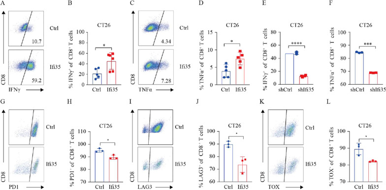
IFI35 promotes CD8+ T cell cytotoxicity and alleviates exhaustion. A, B Representative FACS plots (A) and quantification (B) of IFNγ expression among CD8+ T cells in CT26 tumor. Two tailed t-tests, *P < 0.05. C, D Representative FACS plots (C) and quantification (D) of TNFα expression among CD8+ T cells in CT26 tumor. Two tailed t-tests, *P < 0.05. E, F Effect of tumor-secreted IFI35 protein on CD8+ T cell effector cytokines. Activated mouse CD8+ T cells were cultured in the presence of supernatant from murine colon cancer cells expressing shRNA against IFI35 (shIFI35) and scrambled sequence control (shRNA) for 72 h. IFNγ and TNFα of CD8+ T cells were calculated by Flow cytometric analysis. n = 3. Error bars represent the mean ± SEM. Two tailed t-tests, ***P < 0.001, ****P < 0.0001. G, H Representative FACS plots (G) and quantification (H) of PD1 expression among CD8+ T cells in CT26 tumor. Two tailed t-tests, *P < 0.05. I, J Representative FACS plots (I) and quantification (J) of LAG3 expression among CD8+ T cells in CT26 tumor. Two tailed t-tests, *P < 0.05. K, L Representative FACS plots (K) and quantification (L) of TOX expression among CD8+ T cells in CT26 tumor. Two tailed t-tests, *P < 0.05

