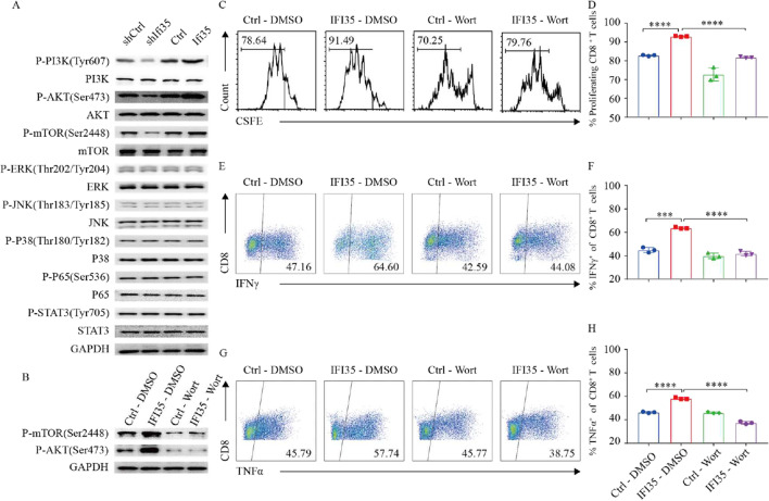
Fig. 7.
Tumor-secreted IFI35 protein activated CD8+ T cells thought PI3K/AKT/mTOR pathway. A Representative Western blots of p-mTOR, mTOR, p-AKT, AKT, p-ERK, ERK, p-JNK, JNK, p-P38, P38, p-P65, P65, p-STAT3, STAT3, and GAPDH in CD8+ T cells. CD8+ T cells stimulated with anti-CD3 (1 µg/mL), anti-CD28 (1 µg/mL), and IL-2 (10 ng/mL) were treated with supernatant from IFI35 knockdown or overexpression CT26 and MC38 cells for 2 days. B, Representative western blots of p-mTOR, p-AKT, and GAPDH in CD8+ T cells. The CD8+ T cells stimulated with anti-CD3 (1 µg/mL), anti-CD28 (1 µg/mL), and IL-2 (10 ng/mL) were pretreated with or without chemical inhibitors Wortmannin (20 nM) at 37 °C for 2 h. C, D The CFSE-labeled mouse CD8+ T cells were pretreated with or without wortmannin (20 nM) for 2 h. The cells were stimulated with anti-CD3 (1 µg/mL), anti-CD28 (1 µg/mL), and IL-2 (10 ng/mL) in supernatant from IFI35 and control vector expressing CT26 for 72 h. Cell divisions were then analyzed by flow cytometry. n = 3. Error bars represent the mean ± SEM. Two tailed t-tests, ***P < 0.001, ****P < 0.0001. E–H Flow cytometric analysis of IFNγ and TNFα of CD8+ T cells. CD8+ T cells were pretreated with or without wortmannin (20 nM) for 2 h and stimulated with anti-CD3 (1 µg/mL), anti-CD28 (1 µg/mL), and IL-2 (10 ng/mL) in supernatant from IFI35 and control vector expressing CT26 for 72 h. n = 3. Error bars represent the mean ± SEM. Two tailed t-tests, ***P < 0.001, ****P < 0.0001

