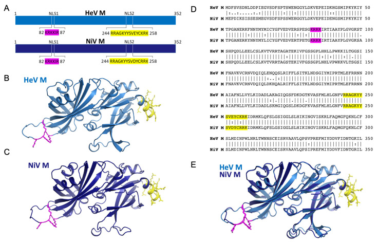
Figure 1.
Henipavirus matrix protein possess two putative NLS regions. (A) Schematic showing the putative NLS regions of HeV M and NiV M. (B) Structure of HeV M in cartoon representation (blue) with the putative monopartite NLS (magenta) and bipartite NLS (yellow) highlighted. The structure was created using alphafold (template PDB70) due to missing loops in the structure of PDB 6BK6. (C) Structure of NiV M in cartoon representation (dark blue) with the putative monopartite NLS (magenta) and bipartite NLS (yellow) highlighted. The structure was created using alphafold (template PDB70) due to missing loops in the structure of PDB 7SKT. (D) Pairwise sequence alignment of HeV and NiV M proteins (E) Pymol image showing superposition of HeV/NiV M from B and C respectively. The RMSD of PDB 6BK6 (HeV M) and 7SKT (NiV M) is 0.57 Å.

