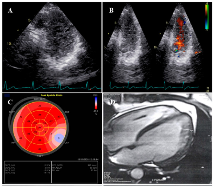
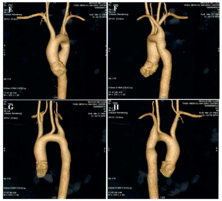
Figure 1.
The imaging results of case #1. (A); Left ventricular apical short-axis view illustrating hypertrabeculated apical portions in addition to deep intertrabecular recesses, (B); Color Doppler echocardiography, showing evidence of direct blood flow from the ventricular cavity into deep intertrabecular recesses (C); Speckle tracking echocardiographic findings, compatible with myocardial performance impairment plus relative apical sparing; GLS = −10.4%. (D); Prominent trabecular network in the apical lateral segments, (E–H); Thoracic CT angiography, showing narrowing of descending aorta, distal to left subclavian artery in different projections.


