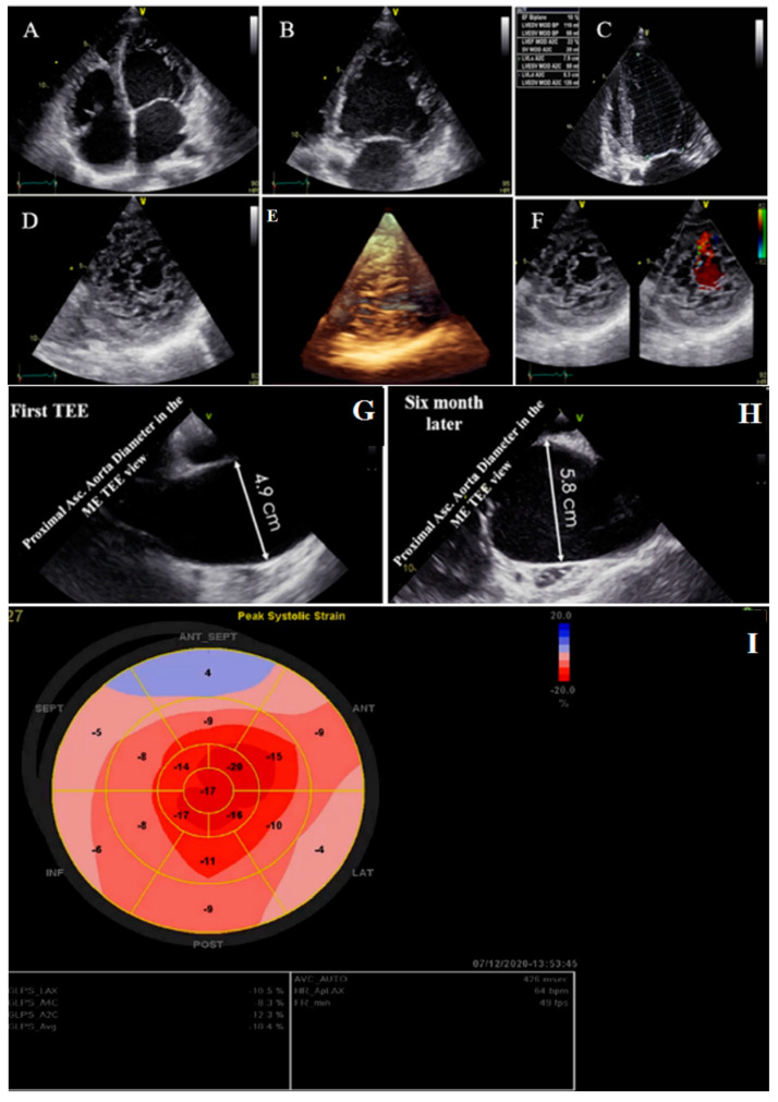
Figure 2.
Two- and three-dimensional transthoracic echocardiographic views of case #2: (A–C); left ventricular apical four and two-chamber views. (D,E); Apical SAX view, illustrating hypertrabeculated apical portions in addition to deep intertrabecular recesses and reduced left ventricular ejection fraction (LVEF = 16%, calculated by Simpson’s method). (F); Color Doppler echocardiography, showing evidence of direct blood flow from the ventricular cavity into deep intertrabecular recesses. (G,H); Transesophageal echocardiographic findings of baseline (left) and 6 months later (right); follow-up test showed fast-growing aortic root that reached 5.8 cm. (I); Speckle tracking echocardiographic findings, compatible with myocardial performance impairment plus relative apical sparing (GLS = −10.4%).

