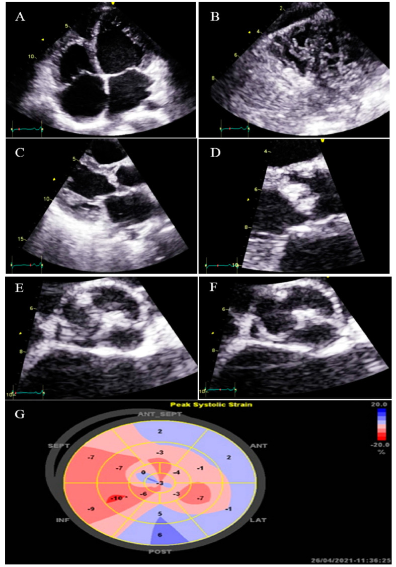
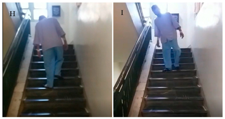
Figure 6.
Two-dimensional transthoracic echocardiographic views of case #6. (A,B); Biventricular non-compaction in apical four-chamber and SAX views. (C,D); Thick, bicuspid aortic valve with diastolic closure doming; PLAX zoomed-out and zoomed-in views. (E); Diastolic closure and systolic opening in SAX view. (F,G); Speckle tracking echocardiography, illustrating severe myocardial performance impairment in all segments with GLS of −3.3%. (H,I); Patient’s difficulty in going up and down the stairs because of weakness in the proximal muscles, when he was asked not to use the stair railing.


