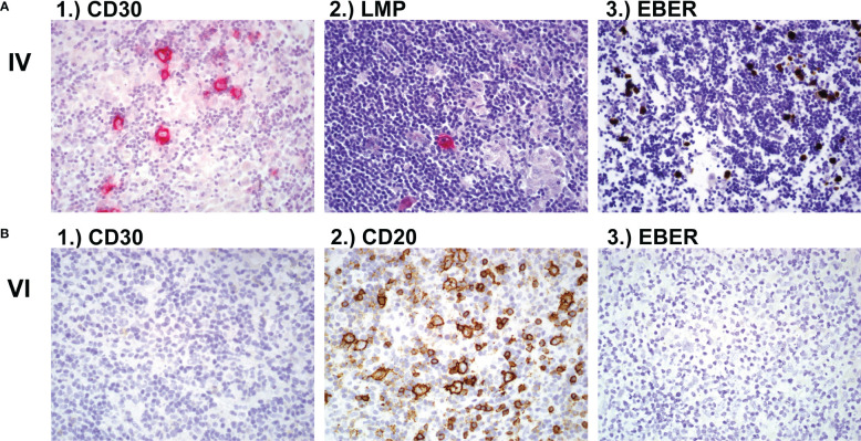
Figure 1.
Lymph node biopsy (A) Lymph node biopsy of family member IV with classic Hodgkin lymphoma showing 1.) CD30 positive cells and 2.) LMP-EBV positive cells after hematoxylin and eosin (H&E) stain and 3.) EBER positive cells after in-situ hybridisation. (B) Lymph node histology of family member VI showing 1.) CD30 negative, 2.) CD20 positive and 3.) EBV negative NLPHL (nodular lymphocyte predominant Hodgkin lymphoma). LMP, latent membrane protein; EBV, Epstein-Barr Virus; EBER, Epstein-Barr virus (EBV)-encoded small RNAs.

