Figure 3.
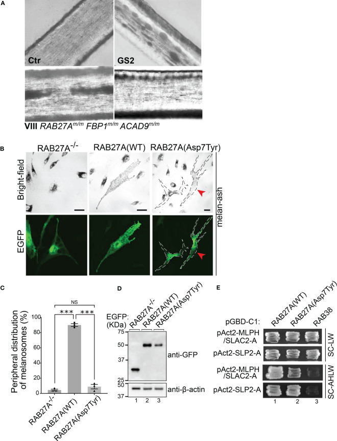
Analysis of RAB27A (Asp7Tyr) effects in hair and cultured melanocytes (A) Light microscopy of hair pigment distribution of a control (Ctr), patient with Griscelli syndrome Type 2 (GS2, RAB27A-deficiency) and patient VIII. (B) Typical images of RAB27A-deficient melan-ash cells transiently expressing EGFP alone, EGFP-RAB27A(WT), or EGFP-RAB27A(Asp7Tyr). EGFP-RAB27A (Asp7Tyr)-expressing cells in the right panels were outlined with dotted lines. Red arrowheads indicate rescued cells with normal peripheral melanosome distribution. Scale bars, 20μm. (C) Percentages of cells showing a peripheral melanosome distribution. Error bars = Mean ± S.E. of three independent experiments (n > 25 cells in each experiment). *** = p < 0.001; NS, not significant (one-way ANOVA and Tukey’s test). (D) Protein expression levels of EGFP, EGFP-RAB27A(WT) and EGFP-RAB27A(Asp7Tyr) in melan-ash cells by immunoblotting with antibodies indicated. (E) Interaction of RAB27A(WT) or RAB27A(Asp7Tyr) with MLPH/SLAC2-A-SHD or SLP2-A-SHD by yeast two-hybrid assays. RAB38, a melanosomal protein that does not bind to RAB27A effectors was used as a negative control (45).

