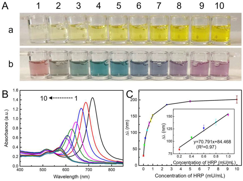
Figure 3.
(A) Optical photographs of produced TMB2+ solutions before (a) and after (b) the addition of Au NBPs. (B) The UV–vis absorption spectra of Au NBPs are etched by various concentrations of TMB2+. From 1–10, concentrations of HRP were 0, 0.1, 0.2, 0.4, 0.6, 0.8, 1.0, 2.5, 5.0, and 10.0 mU/mL, respectively. (C) Plot of Δλ vs. concentration of HRP. Inset: the linear relationship between Δλ and the concentration of HRP from 0.2 to 1.0 mU/mL.

