Figure 8.
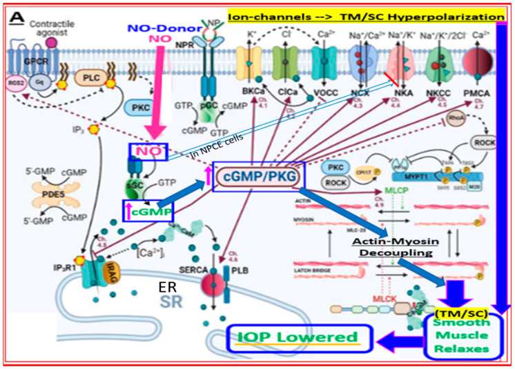
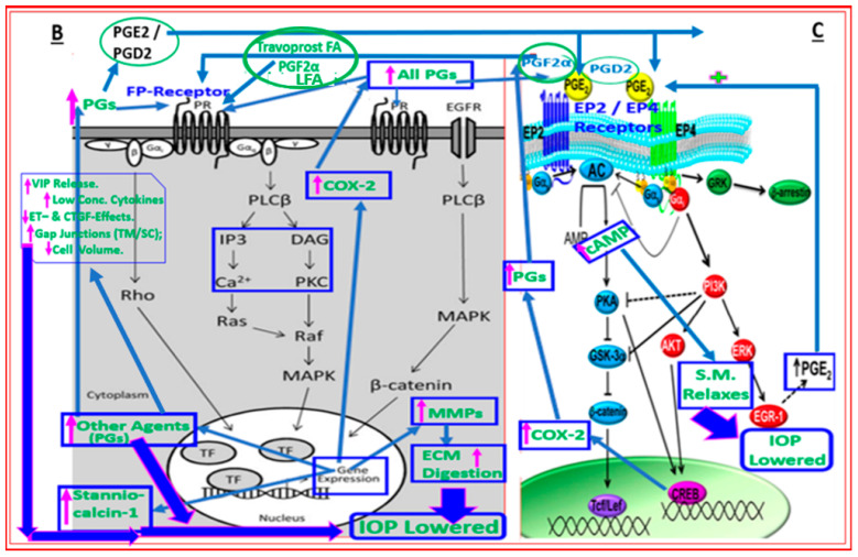
(A) illustrates how nitric oxide (NO), either produced by neighboring endothelial-like TM cells or by NO-donor drugs, elicits signaling in the smooth muscle-like TM cells. Binding of NO to soluble guanylate cyclase generates intracellular cGMP, which activates protein kinase G that uncouples actin-myosin to relax the TM cells, which results in enhanced outflow of AQH from the anterior chamber and thus a reduction in IOP. The activated PKG also reduces the availability of intracellular Ca2+ by inhibiting the endoplasmic reticular calcium transporter and by inhibiting a range of ion channels that hyperpolarize and relax the TM and /or SC cells to lower IOP. Direct inhibition of rho kinase by PKG is also possible. In the case of non-pigmented ciliary epithelial (NPCE) cells, there is evidence that the NO-cGMP-activated PKG can inhibit Na-K-ATPase to reduce AQH production and help reduce IOP (see the light blue outlined and unfilled arrow). (B) shows the various cellular and intracellular components involved in mediating the effects of FP-receptor agonists within smooth muscle cells of the ciliary muscle, TM, and SC to promote efflux of AQH from the anterior chamber of the eye to lower IOP. Here, the phospholipase C activated upon binding of the FP-agonist hydrolyzes cell membrane phospholipids to generate intracellular inositol phosphates and diacyl glycerol, which in turn elicit intracellular Ca2+ release and activation of protein kinase C, respectively, leading to enhanced myosin-activated protein kinase (MAPK) activity. Migration and interactions of MAPK with nuclear materials cause the generation and release of pro-matrix metalloproteinases (MMPs) into the cytoplasm of target cells. The latter are cleaved to liberate and activate MMPs that digest ECM to create or enlarge spaces between CM fibers and around TM to allow AQH outflow via both UVS and TM/SC pathways, thereby lowering IOP. Since cyclooxygenase-2 (COX-2) is also increased, additional prostanoids are generated and released into the extracellular space. These bind to their cognate receptors, and the signal transduction is further amplified (e.g. (B,C)). (C). Numerous other events occur, and endogenous hormones/peptides (e.g., Stanniocalcin-1; vasoactive intestinal peptide (VIP)) are released that also help AQH outflow and stimulate additional IOP-lowering.


