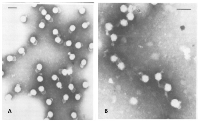
Figure 1.
Earliest electron micrographs of bacteriophage φ6. (A) The bacteriophages isolated from purified lysate, the envelope structure and “saclike” tail are clearly visible. The “saclike” tail is a preparatory artifact. (B) The bacteriophages attached to the pili. The micrographs were used with permission of American Society for Microbiology, from Vidaver et al. 1973 [1]; permission conveyed through Copyright Clearance Center, Inc.

