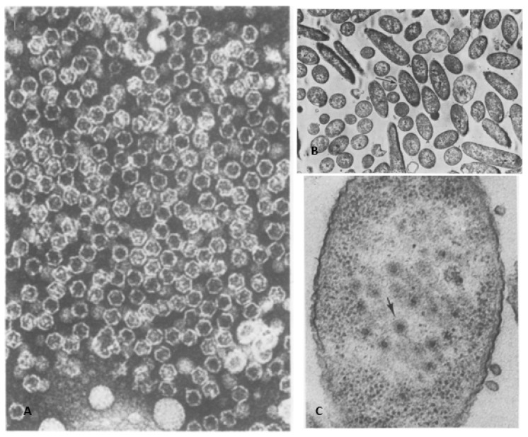
Figure 2.
(A) The first published electron micrograph of chloroform-treated bacteriophages. The icosahedral nucleocapsids were exposed after outer lipid layer removal. (B) A thin section of the φ6 infected P. syringae cells prior to the lysis event, 150 min post infection. The samples were stained with UAc. (C) Magnified central area of the infected bacteria. The arrow indicates the viruses located in the central part of the cell. Images were used with permission of American Society for Microbiology, from Ellis, L.F. and R.A. Schlegel, 1974 [7]; permission conveyed through Copyright Clearance Center, Inc.

