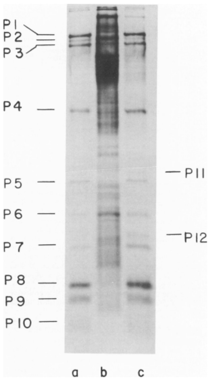
Figure 4.
First SDS-PAGE gel showing the φ6 bacteriophage proteins migration rate and assignment of the proteins according to their motility in a 15% discontinuous polyacrylamide gel. The autoradiograph of the 14C-leucine labeled samples was exposed for 2 days to allow all the bands to appear. (a) Proteins from the purified φ6, (b) uninfected cell lysate, and (c) rifampin-treated φ6 infected cell lysates. Image is used with permission of American Society for Microbiology, from Sinclair, J.F., et al., 1975 [29]; permission conveyed through Copyright Clearance Center, Inc.

