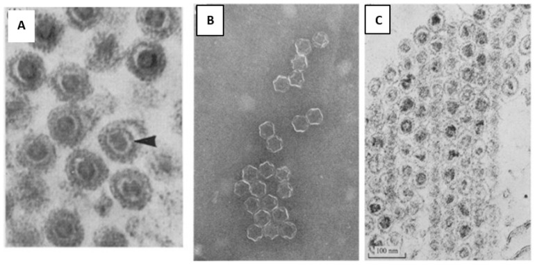
Figure 7.
First attempts of structural analysis of phi6. (A) The bacteriophage thin section, where the different parts of the head of the phage are clearly seen. The outer diameter of the virion is 65 to 75 nm, inner dense core 30 nm in diameter, then there is another electron dense shell ~50 nm in diameter, which appeared as a dark circle on the micrograph and a bi-lipid membrane of ~7.5 nm thickness. The arrow indicates the 50 nm particle, surrounding the 30 nm core. The outermost dark circle is the phage membrane; (B,C) Triton X-100 treated phages, (a) A negatively stained preparation where the 45 to 50 nm large rather complex capsid structure is seen. (b) Sectioned pellet of the phage, where the 30 nm core is seen inside the 50 nm particle. Used with permission of Microbiology Society from Bamford et al, 1976 [43], permission conveyed through Copyright Clearance Center, Inc.

