Fig. 3.
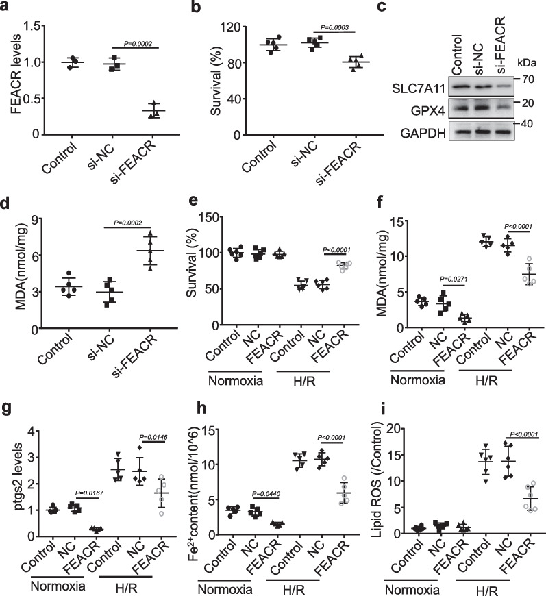
FEACR inhibits H/R-induced ferroptosis in cardiomyocytes. a The expression of FEACR was inhibited in cardiomyocytes measured by qPCR. b CCK-8 kit determines the cell survival rate after FEACR inhibition. c The expression of ferroptosis-related proteins after the knockdown of FEACR was revealed by Western blot. d The content of lipid peroxidation production MDA in cardiomyocytes upon FEACR inhibition. e, f Cell survival rate and MDA content were measured after overexpression of FEACR in cardiomyocytes subjected to H/R treatment. g QPCR analysis measured the ptgs2 level after overexpression of FEACR in H/R-treated cardiomyocytes. h Iron content was determined by ferrous iron kit in cardiomyocytes. i Quantitative statistics of ROS content stained with C11-BODIPY in cardiomyocytes. a n = 3 biological replicates; b n = 5 biological replicates; c n = 3 biological replicates; d, f–h n = 5 biological replicates; e n = 6 biological replicates; i n = 6 biological replicates. Data were mean ± SD, and the P-value was calculated by One-way ANOVA. The experiment technically repeats three times

