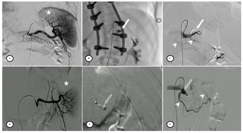
Figure 3.
Preventive proximal splenic artery embolization materials. (A) Upper pole splenic trauma without focal vascular anomaly (star). (B) Penumbra occlusion device (arrow) deployment through a microcatheter along the left lateral aspect of the spine; note that the patient had osteosynthesis material. (C) Final control shows complete flow stasis in the splenic artery downstream from the embolic material (arrow) and the development of collateral circulation (arrowheads). (D) Another upper pole splenic trauma without focal vascular anomaly (star). (E) Amplatzer vascular plug deployment (blank arrow) directly through the Cobra 2 4F catheter. (F) Final control displays the development of a collateral pathway through the dorsal pancreatic artery and the great pancreatic artery (arrowheads).

