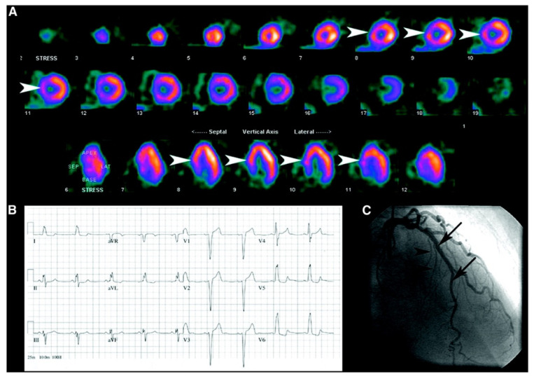
Figure 1.
Patient with chest pain shows septal perfusion defect. (A) Myocardial perfusion scan obtained after injection of 99mTc-sestamibi during episode of pain shows septal defect (arrowheads). (B) Patient’s ECG reveals presence of LBBB. (C) Patient’s coronary angiogram reveals normal vessels supplying the septum, including left anterior descending artery (arrows) and septal perforators (arrowheads), indicating that septal defect was secondary to LBBB [26].

