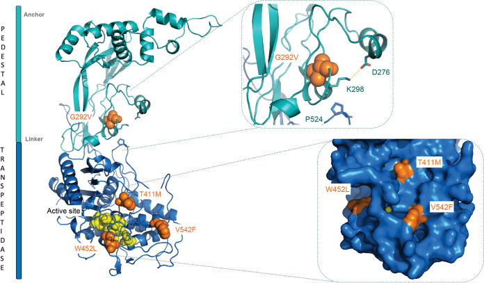
FIG 2.
3D homology model of PBP2b. The pedestal domain is shown in teal, and transpeptidase domain is shown in blue. Amino acid substitutions found in the four wpsH pbp2b mutants are displayed as orange spheres. The catalytic amino acid residues are highlighted as yellow spheres. Inserted at the upper right is a closeup view of the G292V mutation and its interaction sphere, with a highlight on the salt bridge between D276 and K298 and on the interaction between K298 and P524. Inserted at the lower right is a closer view of the accessible surface of the active site that contains the three other substitutions.

