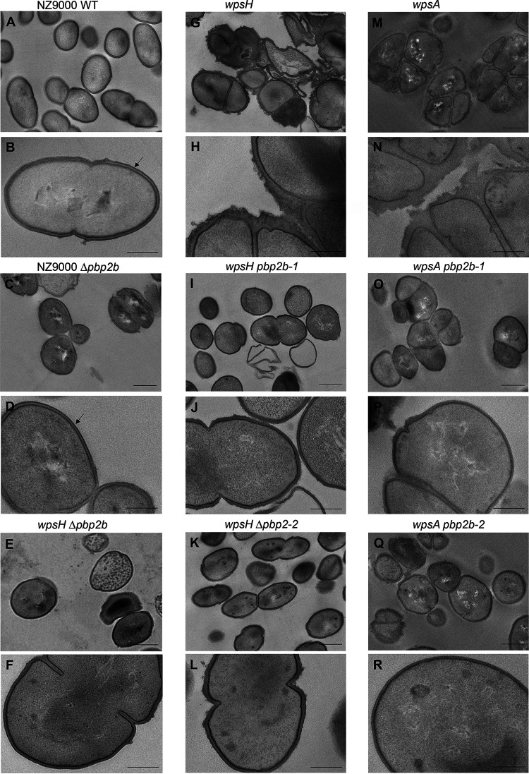
FIG 4.
TEM images of L. cremoris NZ9000, PSP-negative mutants, and their pbp2b derivatives. Black arrows indicate the outer electron dense layer attributed to PSP. Scale bars, 500 nm (A, C, E, G, I, K, M, O, and Q) and 200 nm (B, D, F, H, J, L, N, P, and R). (A and B) NZ9000 WT strain, showing typical ovoid shape, septation at midcell and PSP layer at the cell surface. (C and D) NZ9000 Δpbp2b (VES7552). (E and F) wpsH Δpbp2b (VES7552). (G and H) wpsH (VES5748). (I and J) wpsH pbp2b-1 (VES7484). (K and L) wpsH pbp2b-2 (VES7485). (M and N) wpsA (VES7810). (O and P) wpsA pbp2b-1 (VES7806). (Q and R) wpsA pbp2b-2 (VES7808).

