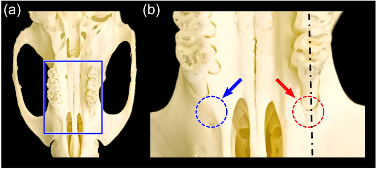
Figure 1.
The three-wall defect of the first molar in SD rats. (a) The surgical field on the rat skull, indicating the selected sample area that was sectioned. (b) Three-wall defect procedures were performed rostral to the upper first molar using a 1.4 mm round tungsten carbide drill. The blue arrow and dotted circle on the left indicate intact bone, while the red arrow and dotted circle on the right indicate the postoperative bone defect, measuring 2 × 1.4 × 1.4 mm³. Note that the dash-dotted line in (b) indicates the histological section.

