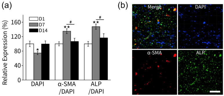
Figure 4.
Immunofluorescence staining of α-SMA and ALP in the collagen scaffolds laden with HPLFs (n = 2 for each 1, 7, 14 day experiment). (a) Relative fluorescence intensity of DAPI, α-SMA/DAPI, and ALP/DAPI at 1, 7, 14 days (n = 6). (b) On the 7th day, both α-SMA and ALP expression were observed, indicating the differentiation of HPLF cells into myofibroblasts and pre-fibroblasts (bar: 100 µm). (* p < 0.05 and ** p < 0.01, compared with D1; # p < 0.05 significant between groups).

