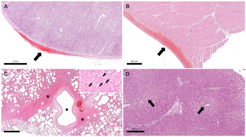
Figure 1.
Donkey, Case 1. Haemorrhagic foci were detected in the renal capsule ((A), black arrow) and epicardium ((B), black arrow). Perivascular haemorrhage was visible in lung along with diffuse hyperaemia and alveolar oedema ((C), black asterisks indicate haemorrhagic foci and black dot indicates an empty blood vessel). At higher magnification, many bacteria were visible in the exudate and in lung alveolar macrophages ((C), black arrows in the image inset). Degeneration of hepatocytes mainly visible in the central part of lobules, near the central veins ((D), black arrows indicate central veins).

