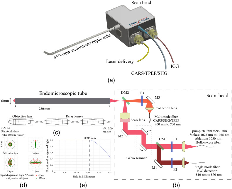
Fig. 1.
(a) Overview of the whole device (excitation laser, PMT detection unit, and scanner controller are not included). (b) Optical path of the system. F1: filter LP750, F2: BP842/56, F3: filter SP750, DM1: dichroic mirror ZT845/42, DM2: dichroic mirror SP749, M1: dielectric coating mirror, M2: silver coating mirror, and M3: silver coating mirror. (c) Optical layout of the endomicroscopic tube. (d) Spot diagram of the endomicroscopic tube at different fields of two wavelengths. Red and green represent 800 nm and 1030 nm. Airy radius is for . (e) Vignetting diagram for field radius from to .

