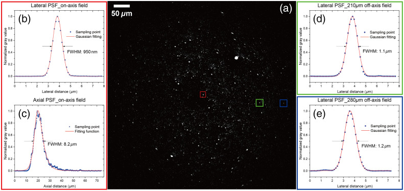
Fig. 5.
(a) 200 nm fluorescence beads (polystyrene, surface modification by carboxylate) image from TPEF signals, excited by a FOPG pulsed laser for multimodal nonlinear imaging (pump beam at 795 nm and Stokes beam at 1030 nm, 5 MHz repetition rate, 22 mW, and 110 mW average power at the sample, respectively for pump and Stokes). Filter: BP514/3. (b), (d), and (e) Lateral PSF of points framed by red, blue, green squares in panel (a), with pixel size of in width, in dwell time, and frame averaged by eight times. (c) Axial PSF of a point at the central field, i.e., point framed by the red square in panel (a). Stack step is in direction. Images were acquired by the ScanImage (Vidrio Technologies).33

