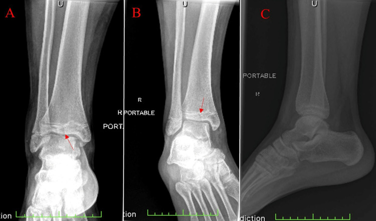
Figure 1. Plain radiographic images from the patient's initial visit to the emergency department.
(A) AP view, (B) internal oblique view, and (C) lateral view. The red arrow indicates the single vertical lucency through the distal tibial epiphysis. Incomplete closure of the tibial physis can be appreciated.
AP: anterior-posterior.

