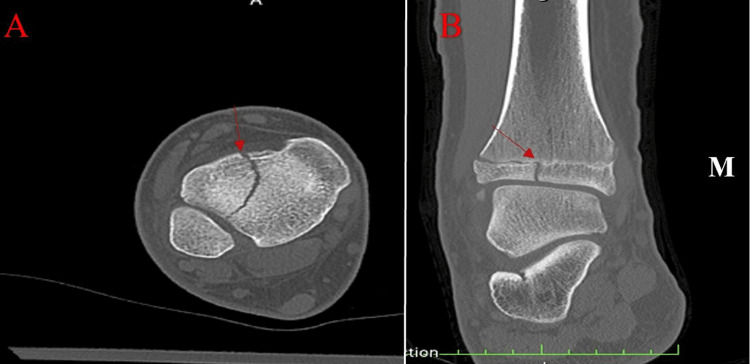
Figure 2. CT scan of the right lower extremity.
(A) An axial view demonstrating the vertical fracture through the anterolateral tibia. (B) A coronal cut of the distal tibia. A red arrow highlights the fracture in both images. Displacement can be visualized on the lateral side, and this fracture pattern is indicative of a Tillaux fracture.

