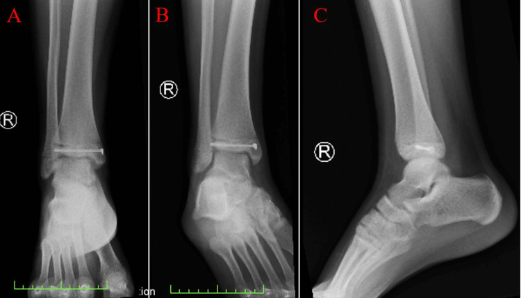
Figure 4. Plain radiographic images post-reduction and fixation of the fracture.
(A) Postoperative AP view, (B) AP oblique with internal rotation, and (C) lateral view. The cannulated screw, reducing the displacement located within the tibia, can be appreciated in each image.
AP: anterior-posterior.

