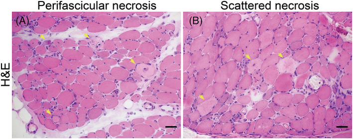
FIGURE 1.

Perifascicular necrosis (PFN): (A) PFN is recognized when two‐third of necrotic fibers present in the two outermost layers at the periphery of the fascicles (perifascicular areas). (B) Scattered necrosis: no specific localization of necrotic fibers. Necrotic fibers are highlighted (yellow arrows) (Bar = 50 μm).
