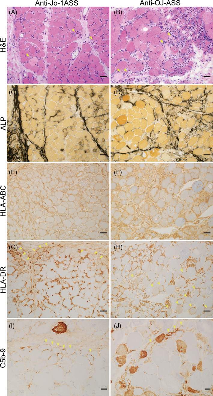
FIGURE 4.

Histologic patterns in anti‐Jo‐1 and anti‐OJ antisynthetase syndrome. Representative figures for anti‐Jo‐1 ASS (A, C, E, G, I) and anti‐OJ ASS (B, D, F, H, J): Both anti‐Jo‐1 and anti‐OJ showed perifascicular necrosis on H&E (yellow arrows indicate necrotic fibers, A and B), but more prominent inflammatory cell infiltration was observed in anti‐OJ ASS (B). Both anti‐Jo‐1 and anti‐OJ ASS showed increased perimysium alkaline phosphatase activity (C and D). HLA‐ABC was diffusely expressed (E and F). Anti‐Jo‐1 ASS was commonly associated with perifascicular HLA‐DR expression (G). In this case, anti‐OJ ASS showed scattered HLA‐DR positivity (H). Both subtypes showed sarcolemmal C5b‐9 expression in perifascicular areas (yellow arrowheads indicate immunohistochemical positive fibers, I and J). A–C, E–G bars = 50 μm; D and H bars = 20 μm; H&E, hematoxylin and eosin; ALP, alkaline phosphatase; HLA‐ABC, human leukocyte antigen‐ABC; HLA‐DR, human leukocyte antigen‐DR; C5b‐9: membrane attack complex.
